

Microscopy
Microscopes cover a wide range of lab, educational, and research needs — from routine sample viewing to fluorescence, confocal, and electron imaging. For the Iran market, current, established, and even archived-but-still-sourceable/serviceable models can all carry commercial value.
Subcategories
Binocular
Binocular light microscopes are among the most widely used lab instruments for viewing tissue, cell, microbiology, and educational samples. The Leica DM750 is an upright binocular microscope for advanced teaching and life-science use, presentable as an established model for the Iran market.
Monitoring
Monitoring microscopes display the image via a camera on a monitor or computer in addition to the eyepiece — useful for documentation, group teaching, and scientific sessions. The Leica DM750/DM500 with a wireless-camera configuration and Wi-Fi solutions serve university labs and teaching classrooms.
Fluorescence
Fluorescence microscopes observe fluorescent structures and markers in biological, cell, and tissue samples. The Leica Visoria B is a current model supporting brightfield, darkfield, phase contrast, DIC, and up to 4-color fluorescence; the Leica DM2500/DM2500 LED is an archived-but-relevant model still useful in pathology and research.
Stereo Microscope
Stereo microscopes provide three-dimensional viewing of larger samples for dissection and educational use. The Leica S9 D is suited to 3D viewing and imaging, and the Leica EZ4 W is an educational model with an integrated Wi-Fi camera for classrooms and universities.
Student & Teacher
Student & teacher / multi-viewing systems let several users observe the same sample at once — used for resident training, pathology, and specialist consultation. Leica Multiviews is a modular solution connecting 2 to 20 viewing stations to one microscope; Leica AirTeach is a digital Wi-Fi alternative for sharing images.
Confocal Microscope
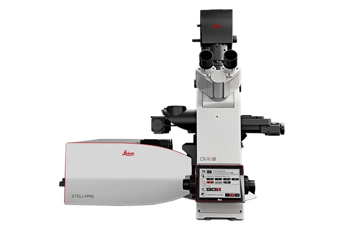
Confocal microscopes use laser scanning and out-of-focus light rejection for layer-by-layer imaging and 3D reconstruction of cell/tissue structures. The Leica STELLARIS platform is the current family and is fully configuration-based — exact detector/laser/objective specs are set only per quote.
Electron
Electron microscopes use an electron beam instead of visible light to observe much smaller-scale structures. The Thermo Scientific Talos L120C is a TEM/STEM with a 20–120 kV voltage range and line resolution down to 0.204 nm, for imaging cells, organelles, and soft materials.
Applications
- Routine viewing of tissue, cell, and microbiology samples
- University and medical education
- Fluorescence and confocal imaging
- Dissection and sample prep with stereo microscopes
- Nanotechnology and materials-science research with electron microscopes